Infrared Spectroscopy
This guide is an early version — the text is complete, and a few figures are still being redrawn. Spotted something unclear? Let us know.
The question this page answers: How can we use information about stretching and bending of bonds to determine FGs?
Deeper reading: Clayden 2e: Chapter 3 Page 63–72 — see our chapter-by-chapter practice map for Clayden.
IR as a diagnostic tool for functional groups
What does IR actually measure?
Infrared (IR) spectroscopy is used as a diagnostic tool for the presence of certain functional groups.
IR radiation in the mid-IR region (4000–400 cm–1) is most useful for organic chemists because it can be used to study the fundamental vibrations of chemical bonds. These vibrations include stretching and bending processes:

Molecules absorb IR energy that matches the energy it takes to vibrate the bond. One caveat: the stretching must induce a change in the dipole of the molecule.

How the IR absorptions of bonds differ
What sets a bond's frequency?
Bonds will vibrate at differing levels of IR energy, which permits us to tell FGs apart.
Here is a generic summary of how the IR absorptions of bonds differ:
- Stretching frequencies are higher than corresponding bending frequencies. (It is easier to bend a bond than to stretch or compress it.)
- Bonds to H have higher stretching frequencies than those to heavier atoms.
- Triple bonds have higher stretching frequencies than corresponding double bonds, which in turn have higher frequencies than single bonds, excluding H.
Here is a slightly more quantitative summary (where A is any atom but H):
- 3600–2700 cm–1A–H (single bond to hydrogen)
- 2700–1900 cm–1A≡A (triple bonds)
- 1900–1500 cm–1A=A (double bonds)
- 1500–500 cm –1A–A (single bonds)
Characteristic peaks of common functional groups
Broad OH, sharp C=O, the 3000 line
Because of H-bonding, IR peaks from ROH and COOH are typically very broad.
The C=O bonds in carbonyl-containing functional groups exhibit sharp IR peaks at different frequencies around 1600–1800 cm–1.
sp2 C–H and sp3 C–H show up on either side of a dividing line at 3000 cm–1.
Triple bonds usually show up around 2050–2250 cm–1.
Amines and amides have N–H stretches in the 3200 cm–1 region.
Terminal alkynes have C–H stretches in the 3300 cm–1 region.
Conjugation and strain shift stretches
Which way do the shifts move?
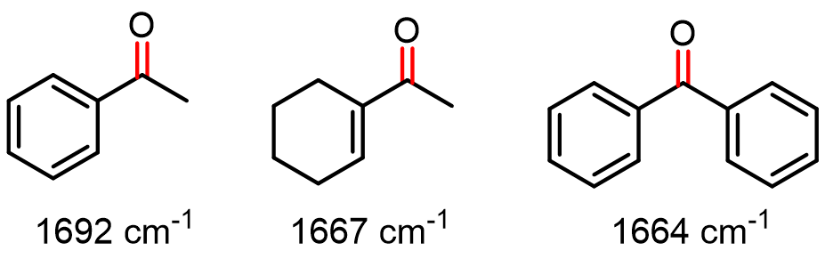
Conjugation shifts stretches to lower wavenumbers. By 30 cm–1 for the first pi-bond.
Strain shifts stretches to higher wavenumbers by ~30 cm–1 per decrease of # of carbons.

Spotted an error, or want a topic covered next? Let us know.